This blog is called ‘Fun With Microbiology’, not’ Fun
With Mycology’ – so what gives?
Why the
disproportionate number of Fungal posts?
Well, this blog came about primarily because, after my
injury, I needed some way to fill my time after regular work hours while
waiting for my ride home.
With my return
to work, I discovered that the laboratory had acquired some new
“Toys” for
photographing interesting microbiological specimens.
Surprisingly, few were interested in
utilizing them, whereas with my previous interest in photography, I was only
too happy to take command of these tools.

This blog is primarily about photography - about describing
microbiology through pictures.
It is
also about “interesting” cases – some of the less commonly encountered observations
usually not described in text books
. (eg Mycobacteria in a gram stain,
Strongyloides tracks on agar plates, VDE
–bacteria that need antibiotics
to survive.) Finally, it is about sharing my photographs with anyone who
may find them of interest.
So, why so much fungus?
Well, bacteria exhibit a limited number morphological variations, as I’ve
attempted to illustrate (right). Generally
speaking, an E.coli cell looks pretty
much like a Salmonella cell, looks
like a Citrobacter cell. While important, yet often subtle, differences
in morphology do occur in bacteria, I have chosen not to pursue them on this
site. Bacteria will be documented when
the topic best lends itself to photographic interpretation.
Generally speaking, there are a limited number of bacterial morphotypes to explore with photography.
While I wish to pursue Parasitology a bit more extensively,
I find myself limited to what cases present themselves in our acute care
community hospital.
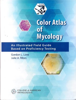
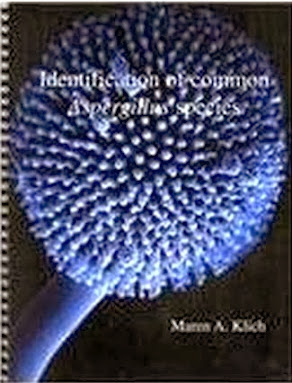
Numerous fine textbooks on Clinical Mycology are in print
(see sidebar); however I have frequently found that the description ‘in text’
relates poorly to the one or two small supporting photographs offered. Fungi are three dimensional organisms whose
structure varies as they mature. I’ve
attempted to document these fascinating organisms from all angles and in all pertinent
stages of development.
In summary, whether saprophytic contaminants or clinically
significant isolates. fungi are the most photogenic microbiological organisms
and present themselves in sufficient numbers to keep this blog going.
Finally, I offer my apologies for the rather lame name of
this Blog. My wife suggested I explore ‘blogging’
as a way to pass the time while bed bound, recovering from a catastrophic injury.
I jokingly chose this name never thinking
I’d develop it past the few print photos I had taken years earlier. Too late to change it now…
 This blog is called ‘Fun With Microbiology’, not’ Fun
With Mycology’ – so what gives? Why the
disproportionate number of Fungal posts?
 This blog is primarily about photography - about describing
microbiology through pictures. It is
also about “interesting” cases – some of the less commonly encountered observations
usually not described in text books. (eg Mycobacteria in a gram stain,
Strongyloides tracks on agar plates, VDE
–bacteria that need antibiotics
to survive.) Finally, it is about sharing my photographs with anyone who
may find them of interest.

.jpg)


